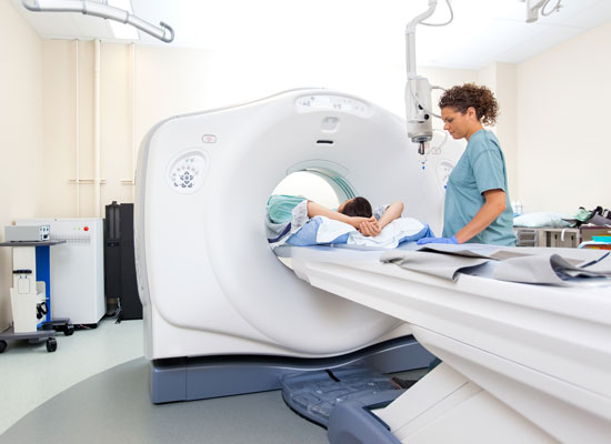
CT Scan facts

- CT scanning adds X-ray images with the aid of a computer to generate cross-sectional view, of anatomy.
- CT scanning can identify normal and abnormal structures and be used to guide procedures.
- CT scanning is painless.
- Iodine-containing contrast material is sometimes used in CT scanning. Patients with a history of allergy to iodine or contrast materials should notify their physicians and radiology staff.
What is a CT scan?
Computerized (or computed) tomography, and often formerly referred to as computerized axial tomography (CAT) scan, is an X-ray procedure that combines many X-ray images with the aid of computer to generate cross-sectional views and, if needed, three-dimensional images of the internal organs and structures of the body. Computerized tomography is more commonly known b its abbreviated names, CT scan or CAT scan. ACT scan is used to define normal and abnormal structures in the body and/or assist in procedures by helping to accurately guide the placement of instruments or treatments.
A large donut-shaped X-ray machine or scanner takes X-ray images at many different angles around the body. These images are processed by a computer to produce cross-sectional picture, of the body. In each of these pictures the body is seen as an X-ray "slice" of the body, which is recorded on a film. This recorded image is called a tomogram. "Computerized axial tomography" refers to the recorded tomogram "sections" at different levels of the body.
Imagine the body as a loaf of bread and you are looking at one end of the loaf. As you remove each slice of bread, you can see the entire surface of that slice from the crust to the center. The body is seen on CT scan slices in a similar fashion from the skin to the central part of the body being examined. When these levels are further "added" together, a three-dimensional picture of a organ or abnormal body structure can be obtained. Than I have given you the list of other specialized procedures.
Scanning Service
INFORMATION - Hispeed N ll Series 7.05 Multi Slice CT Scan. Vipro Ge.company Areb
Certmark AERB CT 28/ 58 - G.S. Dual Ge. Comp.
- CT- BRAIN & ORBIT AXIAL & CORONAL SEC.
- CT- BRAIN + ORBIT + PNS AND ORBIT AXIAL & CORONAL SEC.
- CT - MASTOID TEMPORAL BONE HRCT AXIAL & CORONAL SEC.
- CT- FACE AND NECK + BRAIN
- CT- CERVICAL SPINE + THORATIC SPINE + L.S. SPINE
- CT- THORAX + THORAX (HRCT)
- CT UPPER ABDOMAN
- CT LOVVER ABDOMAN
- CT WHOLE ABDOMAN + CT KUB REGION
- CT NECK AND PNS
- CT- THORAX + UPPER ABDOMAN + NECK + WHOLE ABDOMAN
- CT- PELVIS + HIPS JOINT
- CT JOINT SEGMENT ( ANYONE)
- CT- PNS + TEMPORAH + SELLA ( 152 MM CUT) LIMITED CUT
- CT- PITUITARY CT - SELLA VERY THINCUT
- CT - T.M. JOINT
- CT- GUIDED :- SPECIAL PROCESS
- CT- GUIDED SITE MARKING
- CT-GUIDED ASPIRATION BIOPASY
- CT- SCAN ALL JOIN /WHOLE BODY SUPINE PRONE/ LATERAL OBLI. VIEWS
- 3D CT SCAN :-THREE DIAMENTION CT SCAN
- 3D BUILD MODEL :
- ANGIO
- CT BONE
- CT LUNGS
- CT SOGT
- MPUR
- NAVG
- CF NAVG SMOOTH
- REFORMATION DETAIL
- REFORMATION STANDARD
- 3D BONE WHOLE BODY JOINT VVHOLE BODY BONES BUILD MODAL.
- 3D FACE/ NECK 3D CERVICAL SPINE- 3DI THOROCIC SPINE WITH 3D
- L.S. SPINE WITH 3 D HIPS JOINTS WITH 3 DI BOTH UPPER LIMB WITH 3 D / BOTH LOWER LIMB WITH 3D.
- CT- ABDOMAN (MPR) (MIP)
- CT. GUIDED BIOPSY
- CT 3D NASAL BONE
CT Scan Angiography
- CT SCAN BRAIN ANGIO
- CT SCAN. CAROID ANGIO
- CT SCAN - AORTIC ANGIO CT SCAN PULMONARY- ANGIO
- CT SCAN - ABDOMINAL ANG101 CT SCAN RENAL ANGIO
- CT SCAN - URO ANGIO GRAPHY
- CT SCAN UPPER LIMB ANGIO
- CT SCAN LOWER LIMB ANGIO
- CT SCAN SAINO GRAM
Specially
- CT SCAN VIRSUAL BRONCHOSCOPHY
- CT SCAN COLONO - SCOPHY
- CT SCAN DENTA SCAN
- CT SCAN - LUNGS 3D CT SCAN LUNGS NAVIGATOR
- CT SCAN WHOLE BODY SCANING SERVICE
- CT SCAN GUIDED BIOPSY SCANING SERVICE
- CT SCAN - UROGRAPHY SCANING SERVICE
- CT SCAN - ANGIO RAPHY SCANING SERVICE
- CT SCAN - 3D CT SCANING SERVICE
