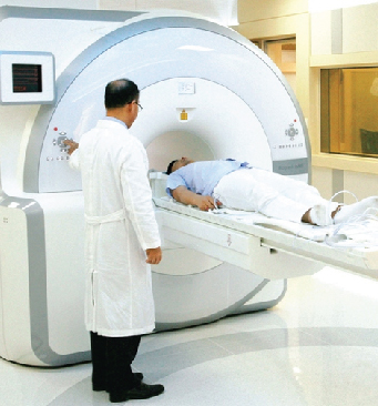


Medicentre Sonography & Clinical Lab, Udaipur, Rajasthan offers a wide range of diagnostic services. The tests & services are available through our imaging, Pathology & Microbiology services. Medicentre regularly adds new diagnostic services and tests.
Medicentre is fully equipped with all latest automated machines in almost all departments to ensure accurate results.To complete the spectrum of services, Medicentre has a number of super speciality labs for Cancer, Neuro, Immuno and other critical diagnosis.
Tests
Satisfied Customers
Years of Experience
Experienced Doctors

Understanding an imaging report can feel like learning a new language. Medical imaging reports (ultrasound and X-ray) are written by radiologists and summarize what the radiologists see, how the exam was performed, and the most important findings (the impression). The impression will be the shor
Read More
Ultrasound In Pregnancy Ultrasound is a safe and non-invasive imaging method that utilizes sound waves to produce images of the growing fetus, placenta, and uterus. It allows physicians to monitor fetal growth, identify abnormalities, and estimate the due date for the baby. Throughout pregnancy, se
Read More
A chest X-ray is a quick, noninvasive imaging exam that uses low doses of ionizing radiation to produce detailed images of your lungs, heart, and chest wall. It's an important tool in diagnosing lung infections, chronic lung disease, enlargement of the heart, and other problems related to breathin
Read More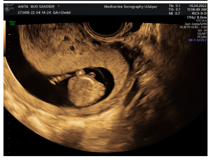
NEURAL TUBE DEFECT (NTD) DIAGNOSED WITH AT 12 WEEKS OF PREGNANCY OF NT SCAN IMAGES SHOW PROTUBERANT ABDOMEN, SHORT SPINE AND LUMBO-SACRAL NEURAL TUBE DEFECT
Read More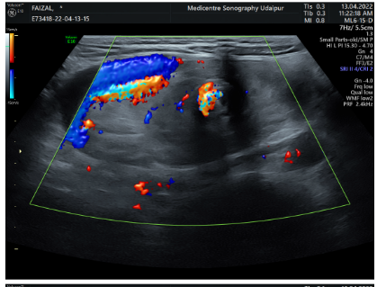
2 YEAR MALE CHILD PRESENTED WITH ACUTE VOMITING FROM 4 DAYS DIAGNOSED WITH PROXIMAL INTESTINAL MALROTATION WITH REVERSAL OF SMA-SMV AXIS ON ROUTINE USG OF ABDOMEN CONFRIMED ON COLOUR DOPPLER
Read More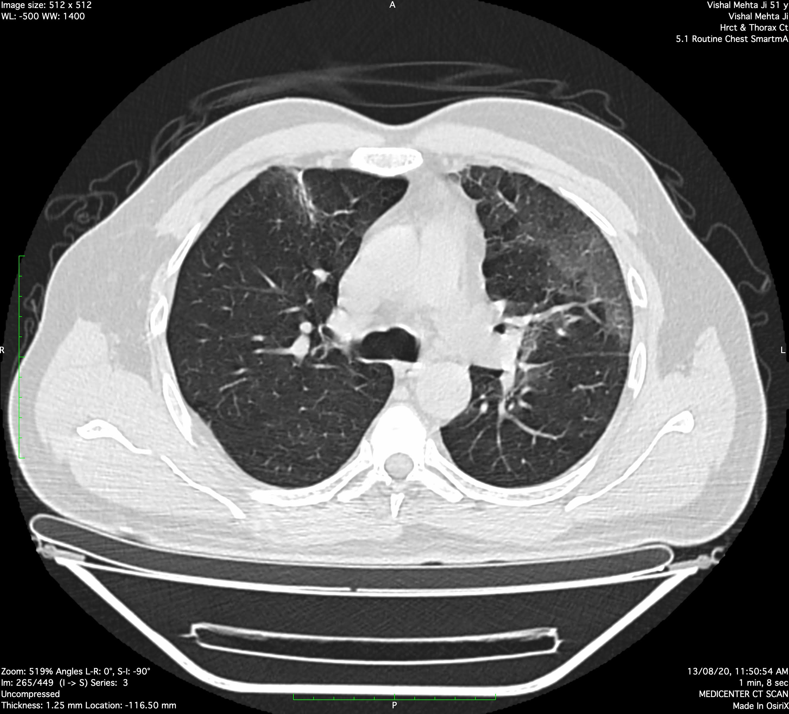
Multiple areas of subpleural and peri-broncho-vascular ground glass opacities with smooth interstitial septal thickening noted in medial and lateral segment of right middle lobe, superior basal segment of right lower lobe, apicoposterior, anterior & inferior lingular segment of left upper lobe, anterior basal, lateral basal and superior basal segment of left lower lobe.
Above findings suggest possibility of atypical pneumonia (intermediate to high suspicion of recent on going pendamic COVID-19 viral etiology). CORADS - 3/4.
Adv:- Clinico-pathological correlation
CT Severity:
Right upper lobe: 0-5 % (1 point)
Right middle lobe: 0-5 % (1 point)
Right lower lobe: 0 % (0 point)
Left upper lobe: 25-50 % (3 point)
Left lower lobe: 0 25 % (2 point)
Total CT severity scoring : 7 points (out of 25).
Read More